Page 31 - Practical-Refraction-English
P. 31
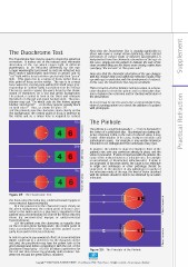
The Duochrome Test Note that the Duochrome Test is equally applicable to those who have a colour vision deficiency; their altered perception of colours (light of different wavelengths) is Supplement The Duochrome Test may be used to check the spherical independent from the chromatic aberration of the eye. In correction. It makes use of the natural axial chromatic this case, simply ask the patient to indicate the side of the aberration of the eye which causes light of different chart on which they see the letters more clearly, rather than wavelengths to be refracted differently by the eye. specifying “the red side” or “the green side”. Longer wavelengths (perceived as red) are refracted less than shorter wavelengths (perceived as green) and so Note also that the chromatic aberration of the eye changes “red” light will be focussed more posteriorly than “green” with the changes that occur within the refractive media of the light. (This gives rise to a range of focus rather than a eye with age; in particular, with the development of cataract. true point of focus on the retina. The eye is in correct In this case, the Duochrome Test may be unreliable. focus when the central point within this small range (cor- responding to “yellow” light) is positioned on the retina). This test can be used for distance and near vision, in a mono- The test is used to assess the eye’s focus by the obser- cular situation to check the sphere and in a binocular situa- vation of characters on a red and green background. tion to balance the correction and for the final verification of The patient is asked to look at the chart and compare the prescription. the letters on the red and green backgrounds. The prac- titioner may ask “On which side do the letters appear At near it may be used to assess the accommodative beha- blacker and clearer? …or do they appear equally black viour of a young patient or to check the addition of a patient on both sides?” Thus, as shown in Figure 28 with presbyopia. a) if the patients sees the characters more clearly on the red background, the central point of focus is anterior to the retina and so a minus lens is required to correct 28a The Pinhole © Essilor International The pinhole is a small hole (usually 1 – 2 mm in diameter) in the centre of a solid black disc. Its principal use during sub- jective refraction is that, in the case of reduced vision, it may enable differentiation of its cause, between refractive and pathological causes. For example, it may enable imprecise refraction to be distinguished from amblyopia (‘lazy eye’). 28b In practice, the pinhole is placed centrally in front of the © Essilor International vision is measured. If vision is improved with the pinhole, the patient’s eye, over any correction already in place, and the cause of the reduced vision is a refractive one; for example an uncorrected, or ill-corrected, refractive error. If vision is not improved or becomes worse, the cause is not refractive in origin and amblyopia or other pathology should be suspected. In the absence of any pathology or opacity of 28c the refractive media of the eye, the level of vision obtained © Essilor International refraction. with the pinhole should be able to be obtained by accurate Figure 28: The Duochrome Test the focus onto the retina (e.g. undercorrected myopia or overcorrected hypermetropia); b) if the patient sees the characters more clearly on © Essilor International the green background, the central point of focus is pos- terior to the retina and so a plus lens is required (or the patient may accommodate) to correct the focus onto the retina (eg overcorrected myopia or undercorrected hypermetropia); c) if the patient sees the characters as equally clear on the red and green backgrounds, the central point of focus is positioned on the retina and the patient is pro- perly focussed for this test distance. In order to prevent any unwanted effects of accommodation © Essilor International (which could lead to a preference for the characters on the red side), the practitioner may have the patient look at the green background before comparing it with the red, or the practitioner may fog by +0.50 D to obtain a preference for Figure 29: The Principle of the Pinhole the red and then remove fog gradually until a balance bet- ween the red and the green sides is obtained. 31 Copyright © 2008 ESSILOR ACADEMY EUROPE, 13 rue Moreau, 75012 Paris, France - All rights reserved – Do not copy or distribute.